Pregnancy is a special new condition of the female organism, when the embryo is beginning to develop in its reproductive organs. With this state sooner or later, you have to face every woman. When it comes to our body, we are looking for a way out and solve medical problems. But when it comes to life two, many women covers panic.
In such delicate and exciting issues, accuracy and confidence are required. Unfortunately, not everything is smooth in our life and have to deal with difficulties. One of these difficulties is the definition of pregnancy. At the present stage of the development of medicine, there are many available and secure methods Pregnancy definitions: diagnostic test strips for the presence of chorionic gonadotropin in the urine, blood test to the same hormone, ultrasound of the small pelvis organs. The difficulty is that these methods can not always be reliable, and should never be relying on one of them.
To determine pregnancy, two ultrasound research techniques are used to monitor the course of pregnancy:
- Transabdominal (Trans Lat. - Through; abdomen lat. - belly) - the sensor is applied directly to the mother's belly to the location of the uterus. Normally, the projection of the uterus is under a pubic symphysome, but during pregnancy the uterus increases and starts to perform more and more on the pubic articulation. The ultrasound directed is reflected from different tissue density and, returning back to the sensor, gives an image of organs in the abdominal cavity. In this case, shows the state of the uterus, giving the opportunity to consider whether it is increased, and see the embryo in her.
- Transvaginal (TRANS Lat. - Higher; Vagina lat.- vagina) - The sensor is introduced through the vagina to a shallow distance, the ultrasound passes through the neck of the uterus, it allows you to consider immediately the organ cavity, bypassing its walls. This method of ultrasound is more accurate, it allows you to consider the absurd of the uterus in a physiological state, the sound does not interfere on other organs and cavities.
Early pregnancy
When it comes to the definition of pregnancy, random or planned, the woman wants to learn as quickly as possible. Ultrasound is not the method that will give an accurate answer in the first days. For this, it is worth contacting other diagnostic techniques that determine the presence of chorionic gonadotropin. Moreover, early timing Pregnancy is not recommended to carry out ultrasound examination without serious readings.

The fact is that in the first days and even the embryo weeks are incredibly tiny. Modern devices ultrasound diagnostics Allowed to see and distinguish between objects in size just a few millimeters, but this is not enough to identify pregnancy in the early stages.
With accuracy, see the embryo on the ultrasound can be 2-4 week of pregnancy. Ultrasound examination in such early dates is carried out only with certain indications that can create a risk of mother's life.
Why is it worth confirming the pregnancy on the ultrasound apparatus?
Always when handling a woman to a gynecologist for registration, it is inspected on the gynecological chair and confirm the pregnancy on the ultrasound of the device. Other research methods are based on the fact that when the embryo appears, there is a change in the hormonal background of a woman, together with it there is a provisional body that allocates a new hormone (chorionic gonadotropin), which tests and capture, confirming the pregnancy.
Ultrasound examination allows you to determine the exact localization of the attachment of the embryo, which allows diagnosing an ectopic pregnancy (fixing the embryo in the fallopian tubes, in the lumen of the abdominal or ovary) and conduct operational interventions in the early periods of pregnancy.
There are also an ultrasound to monitor the development of organs and kids systems, the exclusion of chromosomal pathologies, malformations.
In some cases, the ultrasound may not be detected germin, but the hypertrophy of the mucous membrane is already clearly visible, which can be incorrectly interpreted as a tumor.
Why is it important to determine the ectopic pregnancy on early?
In the early deadlines, an ectopic pregnancy is no different from a normally flowing pregnancy. The chorionic gonadotropin of a person is also registered in the blood and urine, and experts cannot guarantee an ectopic pregnancy, since the fruit egg could not have time to go down from the pipes to the endometrium of the uterus. If after the third week the device does not see pregnancy, then inpatient observation is needed. Launched ectopic pregnancy can seriously threaten the life of a woman.

Bleeding heat, the fall arterial pressure and sharp pain at the bottom of the abdomen - a bright picture of the disturbed out music pregnancy. With progressive ectopic pregnancy, the hazard is high, proceeds asymptomatic, and the size of the uterus corresponds to the norm. But the one, and the other ectopic pregnancy at further flow will lead to the breaking of pipes and extensive bleeding, which will lead to the death of a woman.
Transvaginal ultrasound allows for another 4-5 week (transabdominal for 6 weeks) to diagnose the presence of a fetal egg in the uterus.
Incorrect study for research
In our body, many bodies are placed, which directly closely contact each other. Some of the closely arranged organs are the uterus and the bladder. In the norm, the uterus is deeply in the pelvis, a little behind the bladder, but already the "pregnant" uterus increases in size and opposes it. When carrying out a transabdominal ultrasound, the bladder must be filled. 
Thus, when filling the bladder, the uterus is raised above, increases in size, and its free review opens. With a transvaginal ultrasound examination, the urinary bubble filling does not matter. As a rule, both studies are carried out in turn.
A woman before visiting the doctor for 20-30 minutes should drink about 300-500 ml of water. When filling the bladder, a transabdominal ultrasound is carried out, after that the patient can be released the bladder, and the doctor can start a further survey.
Also, for better ultrasound, it is necessary to control the patient's intestinal state. Gas in the intestinal lumen can be an interference to carry out high-quality ultrasound diagnostics, as the transition of a liquid or solid medium into a gaseous gauge does not let the wave of sound further, and it goes back to the sensor. Therefore, it is impossible to listen to the cavity with the gas field of the body. For 2-3 days, you need to refrain from gas-forming foods. Such are all products containing fiber (vegetables, legumes, especially all kinds of cabbage), sweet fruits enhance fermentation processes (especially grapes), yeast black and white bread, nuts, seeds, carbonated drinks.
Features of the anatomical structure of the uterus
Such individual differences from the usual structure of the uterus can be every person and hide a true pregnancy.
Outdated or defective
Unfortunately, this is the common problem of small cities having a single clinic on the city and often one apparatus. The allocation of a small budget to hospitals sometimes causes specialists to use a half-humor apparatus, which refracts the ultrasound wave, has an uneven frequency, the sensor does not catch the reflected sound, as a result, the image remains distorted.
Unqualified doctor
Even the presence of a convenient, modern and multifunctional apparatus of a new generation does not give a patient a guarantee for accurate diagnostics. An inexperienced employee may not notice a small education or can not cope with the work on the equipment.
As mentioned earlier, one of the signs of pregnancy is the thickening of the inner wall of the uterus to attach the embryo - myometrium hypertrophy. The same picture with the thickening of the wall indicates a benign tumor - the mioma of the uterus. A unskilled doctor may not notice a fruit egg and make a diagnosis of uterine. In the direction of the puncture of education and scraping, a picture opens that "Myoma of the uterus" - normally undergoing pregnancy, but at the moment it becomes too late.
Conclusion
Ultrasound is a modern, safe and fairly accurate method for diagnosing pregnancy. Although the method does not always be absolute, its value is the highest among others, since it reveals not only the presence of pregnancy, but also its possible pathology, allows you to monitor the development of a child in dynamics. For the most part, with falsely unbearable pregnancy, it can be installed in re-ultrasound studies.
For many married couples, there is nothing better than two strips on the test, which give hope for the rapid appearance of the baby. However, the test is not yet an unequivocal positive answer, the gynecologist should also be examined. The most reliable way to learn about my "interesting situation" and remains ultrasound. Another question is on what duration of ultrasound shows a pregnancy.
Ultrasound procedure
To determine pregnancy, two types of ultrasound are used - transvaginal and transabdominal. In the first case, the sensor is used, which is entered into the vagina. In the second - the study is carried out using a special gel. Transabdominal type are mainly prescribed after the first trimester of pregnancy. Such ultrasound gives the overall picture of the state and development of the fetus, and also allows you to see some embryonic pathology.
In small deadlines use transvaginal ultrasound. This method is more reliable and accurate. Thanks to him, experts easily determine the ectopic pregnancy.
After how much the ultrasound shows a pregnancy depends on the day of conception. Mostly ultrasound "sees" a fertilized three-week egg. However, in some cases prescribed a re-ultrasound even in five to six weeks.
Why doesn't the ultrasound show pregnancy?
Ultrasound is a mandatory view of the study during pregnancy. It is used not only to confirm " interesting position", But for the exception of different pathologies. For example, if you do not define an ectopic pregnancy on time, a pipe or ovary can happen (depending on where the egg fixed). However, not always in the early deadlines of the ultrasound of a small pelvis shows pregnancy. This happens for several reasons:
- The fruit egg does not allow to see the physiology of the uterus.
- Failures in the technique.
- Unqualified specialist.
- Very small time (up to three weeks).

This question can be solved by making re-ultrasound seven or ten days. During this time, the egg cell "will grow" and will seem on the screen of the ultrasonic apparatus.
Why does an ectopic pregnancy occur?
Pregnancy outside the uterus - pathology observed in 3-4% of women, in which the fertilized egg is not developing in the uterus, but in other, unaccustomed places - ovaries, uterine tube, abdominal cavity.
It cannot be said that an ectopic pregnancy is a diagnosis of a certain type of women. Such pathology may arise both in healthy patients and in suffering from diseases of the genital tract.

The causes of ectopic pregnancy can be:
- Inflammation of the organs of a small pelvis.
- Failures in the work of the endocrine system.
- The obstruction of phallopy pipes, which arises as a result of infected infections and adhesions.
- Anomalous structure of uterus and appendages.
- Transferred stressful situations.
- Not healthy image Life is the abuse of narcotic substances and alcohol.
- Transferred serious diseases that have given complications for childbearing organs.
The worst consequence of ectopic pregnancy is infertility. However, if pathology in time to determine and eliminate, that is, all the chances of getting pregnant and give birth to a healthy child.
Symptoms pointing to ectopic pregnancy
In small deadlines, an ectopic pregnancy is difficult to diagnose. There is a delay in menstruation, toxicosis, breast augmentation, irritability, drowsiness and some other symptoms. It is possible to determine pathology only with ultrasound. At what period of ultrasound shows pregnancy outside the uterus, depends on the competence of the doctor. Often it is three or four weeks. But sometimes you have to wait up to five to six weeks, which in the presence of pathology can be very dangerous to the health of a woman. Therefore, in addition to ultrasound, it is better to make additional analyzes. In particular, the analysis on the hormone level hCG gives a 100% result. So, when pregnancy proceeds normally, the amount of this hormone is constantly increasing in the body. The weak level of hCG indicates the development of ectopic pregnancy.

In addition to analyzes, the main signs of pathology are also painful sensations at the bottom of the abdomen and allocations resembling menstrual. Often such symptoms are confused with starting miscarriage. Therefore, in such cases it is important to know the exact location location of the fetal egg, so that it is preserved in time to preserve pregnancy, or produce surgical intervention, since the duration of the phallopyye tube can happen on the period of 4-6 weeks.
Operational intervention
Ectopic pregnancy is fraught not only by infertility, but also the death of the patient. Early pregnancy diagnosis helps to avoid such consequences.
If a woman has passed all examinations and suspicions of the doctor are confirmed, it is necessary to operate the patient as quickly as possible and remove the fruit egg. The operation is carried out in two ways:
- Laparoscopy. On the stomach woman is made three small lanes. With the help of special tools, an egg is removed, while the uterine tube remains a whole and able to take part in the fertilization. For laparoscopy, pregnancy period should not exceed 3-4 weeks. If you suspect the ectopic, you should not wait, whether the ultrasound pregnancy will show - it is better to resort to other research methods (HCG).
- Laparotomy. Under general anesthesia, an incision is made on the front wall of the abdominal cavity and the fallopian tube is completely removed along with the egg. Chances of pregnant after such an operation are reduced twice.
Pregnancy planning after ectopic
Although an ectopic pregnancy is an unpleasant experience, but this is not a sentence. On time spent surgery gives a woman a chance to become a mother.

Re-pregnancy should be planned at least in a year - one and a half. During this time, the body will have time to recover.
The whole period before the conception of the doctors is recommended to be protected by oral contraceptives, after the abolition of which the ovaries begin to work "at full capacity" and pregnancy comes almost instantly.
Immediately before conception, it is important to undergo a deep medical examination and pass the necessary assays. So it appears more chances for a healthy uterine pregnancy.
When after a course of therapy and restoration of the body, two stripes are again visible on the test, you should immediately consult a doctor. The re-"interesting situation" makes a woman more awareness, she already knows how the ultrasound shows a pregnancy and how to behave if the small period is not yet visible using ultrasound.
Leaky of normal pregnancy
After a complete examination of the doctor and confirmation of uterine pregnancy comes a responsible moment in life future mom. The first thing to be done is to start leading a healthy lifestyle, without alcohol and harmful substances. It takes more time in the fresh air, to use only useful food and vitamins.
The second important step is to become registered with the gynecologist. It will observe the course of pregnancy, give advice and recommendations that contribute to the birth of a healthy baby.

An important method of diagnosing the development of the fetus is ultrasound. At what period of ultrasound shows pregnancy - depends on the day of conception. If there are no pathologies, a fertilized egg in the uterine cavity is visible on the third week. To eliminate the fret eagle, the doctor may appoint a re-ultrasound on the fifth-sixth week. By this time, the embryo will grow up and will become like a fish.
Further, in the first trimester (10 - 12 weeks), in the second (20 - 24) and the third (34 - 36), an ultrasound is prescribed again to determine normal development. Pregnancy - the norm of development, abnormalities and pathology - is best visible with the help of ultrasound. Therefore, in no case should not ignore this method of research, guided by grandmother's prejudices about the "harm" of such radiation.
Pathology of development of the fetus on ultrasound
Thanks to ultrasonic diagnostics, the pathology of the fetus can already be determined in the early terms and, if possible, to adjust. Some pathology of correction are not amenable, they may be incompatible with life. In this case, the interruption of pregnancy is shown.
So, intrauterine can be determined:

Rely only on some results of the ultrasound. Additionally, analyzes should be carried out, which in combination with ultrasound will give a complete picture.
Conclusion
Pregnancy is a miracle, which are looking forward to many parents. However, so far there are pathology of pregnancy, it can not always be pleasant ...
Does ultrasound ectopic pregnancy and malformations, largely depends on the competence of the doctor. So, planning a "interesting position", you need to choose only an experienced specialist.
Many methods are used to establish pregnancy. Tests that can be purchased in any pharmacy are quite accurate in order to show pregnancy. The same can be said about visiting an obstetrician-gynecologist, which can rather establish the fact of its presence. But it is possible to accurately install it only with the help of ultrasound.
However, it happens that the woman has all the signs of pregnancy: the latency of the menstrual cycle, a positive test and an inspection of an obstetrician gynecologist, which confirms the presence of a pregnant woman, and ultrasound does not define pregnancy. Further, let's try to figure out if it can be that ultrasound does not show pregnancy, in the event of a delay of menstruation and positive test.
Why ultrasound doesn't always show pregnancy
Ultrasound examination is not appointed just like that. Most often, the reason is to inspect the gynecologist and receiving two or more positive pregnancy tests. After all, it is rather strange to come to ultrasound without having any signs, but only suggesting pregnancy, after the presumptive conception.
But, of course, the option of self-evaluation into the office of ultrasound diagnostics is not excluded, if there are symptoms that are pushing on the pregnancy, namely:
- sharp change of emotional background;
- pulling pain at the bottom of the abdomen;
- nausea;
- change of appetite;
- weakness.
However, and with all the above "symptoms" and positive test, ultrasound may not show pregnancy.
Cases when ultrasound does not show pregnancy when delayed menstruation
- The first symptom of pregnancy, which many centuries relied on people, not even having the possibility of accurate diagnostics - delay. However, its cause can be many diseases. First of all - violations hormonal background. These can lead tumors, stress, endocrine system pathology. Sometimes it can be associated with moving to another climatic belt, and sometimes at all due to a sharp change of weather.
- Another reason is exhaustion. The girls who wear out their body can also bring to the point that the menstrual cycle will not come. But still, the most dangerous - malignant tumor diseases, because, if, a woman will neglect the campaign to the Cabinet of Uzi and will assume that it is pregnant - it may well lead to very poor consequences, especially due to tumors, the test can also be positive .
It is worth understanding that the above cases may appear from any girl against the background of absolute health, and she herself often does not notice any changes in their health, continuing to live, not suspecting anything.
In which cases ultrasound does not show a pregnancy in early terms
 However, even if all pathologies are excluded, then not everything is so simple. In small deadlines, it is quite realistic to see pregnancy. Starting from 5 days of delay, which will be approximately equal. The ultrasonic apparatus can show the presence of a fetal egg, but a little later, the same ultrasound device may not determine the pregnancy. From this it should be concluded - you should not hurry.
- The reason for the absence of pregnancy in the ultrasound can be a banal miscalculation in the periods of menstruation and delay. Because of this, the fruit egg is not detected, since at the moment of diagnosis, it is most likely in, and not in.
- Ultrasound during pregnancy in the early terms is important and due to the occurrence of ectopic pregnancy, which can not be seen. Associated, such problems, with a feature attaching a fetal egg in anomalous places and its small sizes. Therefore, in early ultrasound studies, preference is given to a transvaginal method, because it is at different times more accurate.
Do not forget about the peculiarities of the devices and the human factor:
- Old ultrasound devices are much less sensitive, and therefore, they will be able to diagnose much later, and above.
- As for the human factor, everything is more complicated here. An inexperienced specialist or one who has not previously dealt with obstetrics and gynecology, can confuse a fruit egg with a tumor and vice versa. Therefore, it is worth carefully choosing the venue of the ultrasound and clarify information about the specialist who will be diagnosed. Because of this, of course, you should not completely trust the first ultrasound studies, as they are erroneous. It is often possible to determine pregnancy due to inflammatory processes, because inflammation leads to a edema, behind which the fetal egg simply may not be visible.
Ultrasound and additional analyzes and tests for determining pregnancy when delay
 After the appearance of a delay, women tend to turn to pregnancy test. The test is sensitive to the growth of the hormone, which increases in the presence of pregnancy. It is called chorionic gonadotropin (HCG). This hormone appears from the first hours of pregnancy, but some tests will not be able to identify it very early, it all depends on sensitivity.
When the ultrasound does not show a pregnancy (a fruit egg), then believing is worth the test, of course, if the hormone-producing tumor is excluded. If it is presented, the level of hCG in non-empty women will be elevated.
In the future, the blood test should be performed to increase the concentration in it hCG. Thus, the woman should understand that it is impossible to estimate the presence or absence of a fetus only with the help of one method.
- The presence of a steadily increasing level of chorionic gonadotropin suggests that pregnancy occurred and proceeds normally, at least in early terms. By the 7-11 week of pregnancy, the level of hCG increases several thousand times, but then its number gradually decreases. With the help of controlling chorionic gonadotropin, in most cases, we can talk about the presence or absence of fetus development. However, the level of this hormone may increase in the case of ectopic pregnancy. This is once again indicating the need for additional ultrasound diagnostics and consultation of a specialist.
- Sometimes biochemical pregnancy can occur. With her, the fruit egg is discouted as soon as it fixed in the uterus. It happens on the first two weeks of pregnancy, and therefore doctors on the ultrasound will not be able to detect it, and the test, often does not show its presence. The difference from the miscarriage is that with a biochemical pregnancy, the presence of a fetal egg is confirmed, that is, in fact pregnancy did not come, unlike the situation with miscarriage, when, before interruption occurred, the pregnancy was stated.
- The reasons why the test and ultrasound does not show pregnancy - a lot. It may simply be the lack of a pregnancy or the death of a fetal egg for some reason. Also, diagnostic methods may not show pregnancy, if the term is extremely small and is equal to several days.
- To differentiate the causes of GGH growth, a woman will need to pass blood for analysis several times, and then re-execute ultrasound to determine pregnancy, possibly in another medical institution. During this period, the doctor will evaluate the presence or absence of growth in the level of this hormone and it corresponds to the norm.
Because of all the above situations, doctors advise not to rush to the diagnosis of pregnancy. Especially when its term does not exceed even 3 weeks. The diagnosis of pregnancy is not a very urgent procedure, because the higher the time, the more accurate will be the result of the diagnosis. But also with a visit to the doctor and the ultrasound of the office should not be delayed, because it was previously indicated, a number of dangerous diseases may hide behind it.
How soonen can you determine the pregnancy by the Uzi method and how many times can you do it?
Most women who suggest pregnancy, want to know - on what time the ultrasound shows a pregnancy? Ultrasonic diagnostic methods allow you to determine its presence for about 3 weeks, but it turns out not always. Many factors affecting the likelihood of detection of the fetal egg, indicated above, but there is another important item - the place where the fetal egg is attached.
- Especially it plays a big role in the presence of.
- Execution ultrasound to determine pregnancy in the early deadlines only in the case of symptoms indicating the presence of pathology that defines an obstetrician-gynecologist. Usually this ultrasound is performed for 7-8 weeks. It can be detected by an ectopic, fromy pregnancy, tumor, etc. To conduct an ultrasound in earlier deadlines, without testimony, it is simply inappropriate, but it will not harm it to the fetus.
- It is a frequent question: "How many times can you do an ultrasound during pregnancy?". Since sometimes it is not visible, ultrasound can designate a newly and woman, worrying about their future child, will experience concerns about the security of such a diagnosis. All ultrasound methods are absolutely safe, since the ultrasonic waves used as radiation are not harmful, neither for the mother nor for its future child.
Based on this, ultrasound can be performed as many times as it takes.
What types of ultrasound to determine pregnancy apply and prepare for them
 The definition of pregnancy is performed by two main methods:
- Performed by the introduction of the sensor in the vagina. This type of ultrasound is most often applied in earlier pregnancy and guarantees higher accuracy of the results. Preparations for transvaginal ultrasound is not required, the only thing is the need to devastate before the study and fulfill the toilet of the external genital organs as in the gynecological inspection . It will take a special, but it is necessary to purchase it yourself or not - the doctor who appoints or conducts a study. Normally, the procedure is painless and takes no more than 10-30 minutes.
- It is carried out through the front abdominal wall and recommended at later deadlines when, somewhere from 5-6 weeks. In the early dates, the preparation will be necessary. It consists in excluding the use of gas-forming products per day before the study, and then, before the study itself, it will be necessary to fill the bladder. This is done to improve the passage of ultrasound waves. On more high timing, preparing is not needed, as the fruit is great enough and there are oil flooded waterwhich are a good conductor.
conclusions
- When a woman has such signs of pregnancy as a change in emotional background, appetite, taste preferences, nausea, fatigue, weakness and of course delay, and ultrasound does not show pregnancy, does not mean that there is no pregnancy. Here you should refer to other methods. First of all, the test, if it is positive more than twice, then this is a completely significant argument, which has a lot of weight, rather than ultrasound. It is no less important to visit an obstetrician gynecologist who will inspect and confirm the pregnancy or will suspect any pathology.
- Another important study is an analysis for the presence of chorionic gonadotropin. Regular analysis will indicate the development of pregnancy or its fading.
- Ultrasound examination can detect hormoneproproduction tumors, the only sign of which is a false positive result of the analysis of hCG and signs of pregnancy.
- Another pathology requiring urgent intervention is an ectopic pregnancy. If it, for example, is localized in the uterine tube, then it may cause it to rupture the pipe, which is very dangerous to life status. In the future, a woman may have problems in trying to get pregnant.
- It is worth understanding that one ultrasound, on which pathology was discovered not enough. And a few weeks later, if the condition does not require urgent surgery, it will be necessary to repeat the examination.
- No need to worry about the safety of ultrasound methods. They are all absolutely safe and do not carry any harm to the body of the mother or child, which is confirmed by the experience of using ultrasound long in more than half a century.
Ultrasound diagnostics To determine pregnancy - a very important diagnostic event, ignore which is extremely not recommended, because only such a method in many cases does not simply detects pregnancy, but also saves life, while not exposing danger or future mother, nor her baby .2 estimates, average: 5,00 out of 5)
Suspicions of a possible "interesting situation" may appear in a woman long before the next menstruation delay. Modern test strips can determine the content of the specific hong hong in the urine on the first day of the delay, and some even a few days before it. Whatever the test result, the woman wants to make sure the pregnancy as early as possible. About when the baby can be first seen on the ultrasound, will be told in this article.

Minimum definition time
After the conception was held, intensive processes begin in the future mother, which she often does not recognize. In the very first day, the fertilized egg is divided and moved along the uterine tube, where the conception took place, in the uterus cavity. Travel it lasts about four days. The uterus is no longer a set of individual cells, but blastocyte - form form in the form of a ball. It is embedded in the uterus shell. This is implantation. It happens 6-7 days after fertilization, and sometimes a woman feels an implantation on a small pulling sensation at the bottom of the abdomen.

Most. early symptom Pregnancy sometimes is the so-called implantation bleeding - a few drops of bloody or succinous discharge at the time of the introduction of blastocytes in the endometrium. This does not mean that it is time to run out of the dough or sign up for an ultrasound.
Test strips react to the formation of the so-called pregnancy hormone - hgch, and it only begins, the hormone level is lower than the test level of the test strip. And on ultrasound blastocyte to see it is impossible - its size is only 0.2 mm.

How is the ultrasound?
To determine pregnancy, two types of ultrasound examination are used - tranny and transabdomomominal. In the first case, the doctor conducts an inspection of the uterus and its contents with a vaginal sensor. In the second case, the inspection is carried out by the sensor through the abdominal wall. Most, doctors prefer the first method, if it comes to early gestation. Through the vagina, it is much easier to see the embryo and its structure.
Ultrasound of the small pelvic organs abdominal methods are recommended to be carried out with full bladder, Transvaginal - with empty, at the same time it is better to take care of the intestine to be not broken from gases. For this, a few hours before the campaign to the doctor, it is desirable to accept "Espumizan" or "Smekt".


It should be noted that a transvaginal pregnancy can be seen earlier than a transabdominal, for several days. So, a vaginal sensor and a good specialist in an impression can tell a woman about her "interesting position" for 5-6 days from the date of delay, and scanning through the stomach may not show pregnancy and 8-10 days. The procedure is painless, non-hazardous for a woman and baby, it lasts not more than 5-7 minutes.

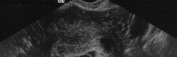
Decoding of the first ultrasound
At the very first ultrasound study, echogenic education will be able to detect the diagnostics. This is a fruit egg. Its dimensions will indicate the exact period of pregnancy. The doctor will also determine the size of the yolk bag, the position of the fetal egg, the thickness of the endometrium, will exclude the inflammatory processes in it, as well as the presence of cysts, polyps and other unwanted formations. The dimensions of the fetal egg and the timeline table are presented below.

Are errors possible?
The method of ultrasound diagnostics is considered one of the most accurate to determine pregnancy in the early stages, but should not be assumed that its accuracy is 100%. In gynecology, the accuracy of this study is estimated at about 90%. In the early period of pregnancy, accuracy decreases to 75%. The doctor is first of all a person, not a machine with the program laid in it. He has the right to make a mistake, especially if a woman has problems with the health of the reproductive system. So, the doctor can confuse Mioma uterus with pregnancy in the initial periods, if earlier the woman was not diagnosed by Mioma, and she found out about it only on ultrasound. A cyst or polyp can be confused with a fruit egg, since the cyst is also an echogenic education.

If the woman had a late ovulation, then pregnancy a week after the delay may not be found in a specialist of ultrasound diagnostics, since the fruit egg later dropped into the uterus and is not yet visualized. Naturally, the doctor will write in the conclusion that there are no signs of pregnancy, but after 7-10 days on a repeated study, it will be able to determine the fruit egg, and its structure. Only the sizes will help to understand that ovulation is really late.

Common questions
In the Internet, inexperienced pregnant and those who still dream of a "interesting situation" ask a lot of questions regarding the earliest diagnosis. About the most frequent situations worth telling more detail.
Pregnancy test gave a positive result, and ultrasound - no
There may be several reasons for this. First of all, it is not worth excluding that the test was defective, it happens, and quite often, especially if we are talking about inexpensive test strips that are sold almost every corner. In the desire to see two cherished stripes, some ladies come too far, starting to look around on the strips of the strip-ghosts. If you find, you automatically begin to consider your test positive, although in reality pregnancy may not be.
If the test still did not deceive, then the reason for the negative conclusion of the ultrasound diagnostic physician may be that the woman turned too early to the doctor, and the fruit egg is not yet visible.. The device itself may be obsolete, with low sensitivity and poor resolution. The reason for the absence of signs of pregnancy on ultrasound may be the late ovulation, and the presence of an inflammatory process in the uterine cavity, and, of course, the insufficient qualifications of the doctor.

Pregnancy test gave a negative result, and ultrasound is positive
The reasons for such a situation may also be enough. First, the test at home the woman could have been done with a mistake, the test could be defective or overdue, and it is also possible that he spent it too early when the hong hong level in the urine was still insufficient, so that the test could respond to it bright second strip.
Ultrasonic diagnosis in this case is rarely premature, since a woman after a negative home test is in no hurry to the doctor, patiently waiting for the beginning of late menstruation. After one and a half or two weeks of delayWhen the lady still appeals to the doctor, pregnancy on ultrasound is already clearly visible. Therefore, the results of the ultrasound should be considered more reliable than the results of the home test. In doubtful cases, you can hand over the HCG to get even more accurate data.

How to calculate the term of pregnancy on ultrasound?
To do this, you can use the table below. If a large periodization of the period is required, use the table matching table up to a day to the average internal diameter of the fruit egg (SVD). Table of pregnancy time in accordance with SVD is shown below.
The value of the average inner diameter of the fruit egg | Gestational age |